Version HTML de base


48
absence of such a fatigue or endurance limit is common in
engineering materials that are either highly defective, e.g.
filled with small defects or that are tested in chemically corro-
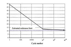
sive environments (fatigue corrosion). Figure 3 described
the behaviour of fatigue accordingly to Carter studies for a
compressive loading of human cortical bones, with an esti-
mated fatigue limit.
Fig. 3 : Fatigue curve for a compressive loading
of human cortical bones [43]
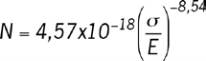
Fatigue behaviour of trabecular bone
The fatigue of vertebrae depends on the trabecular bone
behaviour. A lot of studies have shown that the fatigue
behaviour of trabecular bone is similar to the behaviour of
the cortical bone. The trabecular bones present a Young’s
modulus of 20% less than the cortical bone modulus and
the S-N fatigue curves have similar slopes, but with lower
stresses. Haddock et al [44] tested 35 cores of fresh and
frozen elderly human vertebral trabecular bones, extracted
from nine donors (ages from 37 years to 97 years old).
The tests were biomechanically conducted in compres-
sion. A relationship was derived between the number of
cycles before damage N and the applied stresses
σ
(MPa),
with a coefficient of determination R
2
of 54%.
(2)
where E ist the Young modulus (MPa).
Fatigue behaviour of cartilage endplates
The endplates are thin layers of hyaline cartilage that cover
the central region of the vertebral body endplates on the disc.
Physically, this tissue is similar to articular cartilage near its
junction with bone, but unlike articular cartilage, it is only
loosely bonded to the underlying bone presumably because
it is always pressed up against the bone by the hydrostatic
pressure of the nucleus. Hyaline cartilage is a connective
tissue with an abundant extracellular matrix that combines
the properties of toughness and compressive strength. It has
a sparse population of cells (chondrocytes) but contains no
blood vessels or nerve endings. It provides a low-friction and
low-wear bearing surface, and is able to distribute loading
evenly on the underlying bone. Vertebral body endplates are
usually flat in young adults, but develop a marked concavity
with increasing age, and this may be indicative of repeated
minor injuries to the endplates themselves or to the vertically
oriented trabeculae which support them. Although fatigue has
been implicated in cartilage failure, few studies can be found
in literature. One research [45] tested articular cartilage along
the perpendicular direction. This type of test corresponds
perfectly with loading in endplates of vertebrae exposed to
vertical vibration. This study investigated cartilage responses
to the fatigue cyclic tensile loading, applied under physiologi-
cal conditions. In this study, only one human knee, 48 years
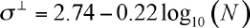
old, has been used. The degree of cartilage degeneration
was visually assessed to single out fibrillated regions. A loga-
rithm relation between the applied tensile stresses
σ
┴
(MPa)
and number of cycles to failure N has been established. This
relationship can be expressed as:
(3)
where
σ
┴
is the perpendicular tensile stress to the colla-
gen fibres. The number of load cycles to failure N thus
varied from 20 to 1.5 10
6
cycles in a range of stresses
varying between 1 and 3 Mpa. In order to consider the
ageing effect on the cartilage fatigue, a similar approach
to Weigthman’model [46] has been used. The fatigue test
has been conducted in tension in the same direction than
the collagen fibres of the articular cartilage. A relationship
between the age Y (years), the stress
σ
┴
and the number
of cycles to injury N has thus been derived:
(4)
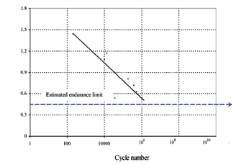
Fatigue behaviour of intervertebral disc
Adams et al [27] have conducted tests by combining bend-
ing and compression loadings at the frequency of 0.67 Hz
during 6 hours (14 400 cycles). From 29 specimens (aver-
age age of 35 years old), 6 have presented radial cracks
conducting to a slipped disk under a load of 3500 N. They
have observed radial cracks in the ring with a degeneration
of the disc. From these tests, the following fatigue curve
can be extracted (Figure 4).
Fig. 4 : Fatigue curve of intervertebral disc
Results
Dynamic analysis
The dynamic behaviour of vertebral bones may depend on
several variables such as: age, sex, posture, loading, excitation
frequency and several other factors. In this study, we consid-
ered the principal following parameters: the posture (
θ
), the
body weight (M), the bone structure (S), the vibratory level (A)
the frequency of excitation (f) and the damping rate (
ξ
). If it is